Skin dose map issues for North Bristol Trust

Ingrid Turner
Good morning,
I am a medical physicist at University Hospitals Bristol & Weston NHS Foundation Trust (UHBW) and our department also looks after the medical physics at North Bristol Trust. North Bristol have acquired OpenREM recently and I have been doing some studies for the skin dose calculations that it does and comparing them to our current method of estimating skin doses. So far this has been for fluoroscopy only as this is where we often get high skin doses that need checking.
I have been studying some skin dose cases for the four interventional radiology labs (Brunel IR1 – IR4) as well as the Hybrid lab at Southmead Hospital. I have found that there are a few issues with the IR4 skin doses as well as the Hybrid lab, and I was wondering if anything could be done with the code for these.
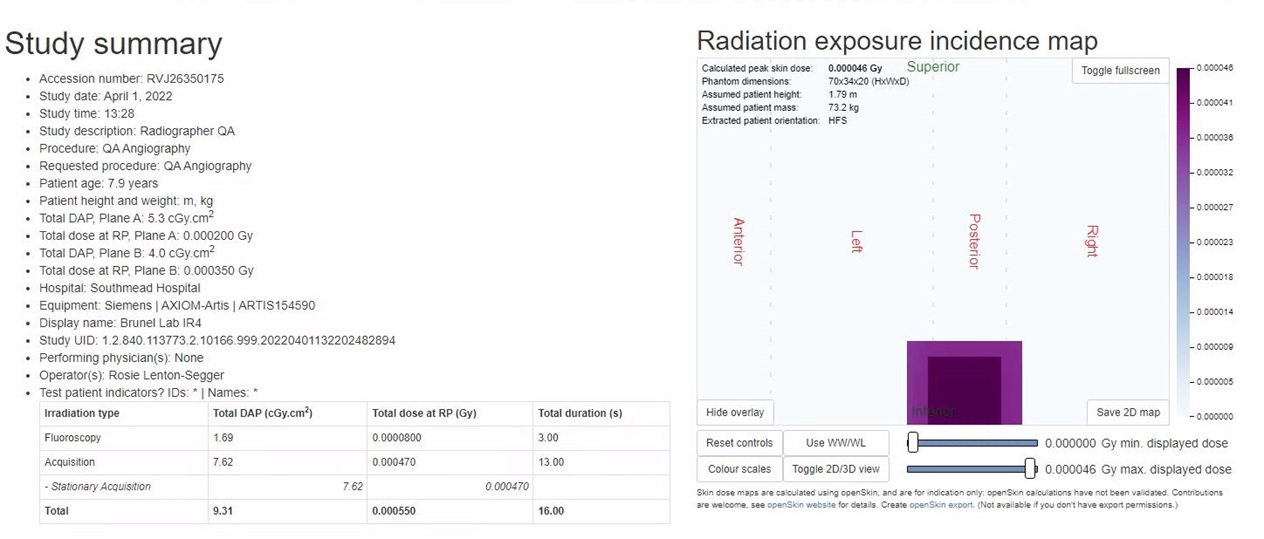
Brunel IR4 is a neuro room with bi-plane imaging – I have found that consistently the skin dose map in OpenREM appears to “fall off” the edge of the phantom and therefore the peak skin doses are being calculated as too low – please see an attached example where the dose at reference point is several Gy but the peak skin dose is far smaller. I have been in consultation with the radiographers for this room who say that a head extension plate is routinely used for neuro examinations. I also asked them to image both ends of the table at full length and I can confirm that the head-end dose does not appear on the phantom (also attached).
For the Hybrid lab, a different length table is used as it is also an operating theatre, and I think this is causing similar problems in openREM, as the dose map consistently appears in the wrong place on the patient – e.g. a kidney examination appears in the head area of the phantom, see attached. Similarly to IR4 I asked the radiographers to image both ends of the table, and nothing appeared on the dose map.
I’m not sure if any adjustment can be made in the code for these two rooms to account for the different table lengths?
Finally a small point – I was wondering if there was a way of inputting the patient height and mass after the initial skin dose map has been calculated? For some examinations it seems this information has not been entered by the radiographers before-hand - normally they give us this information separately when they want us to look at a high skin dose case. I read in the documentation that this can be done but I could not find an obvious way of doing this?
Apologies for the long email, I hope this all makes sense and I look forward to hearing from you.
Many thanks and best wishes,
Ingrid
*************************************************
Ingrid Turner (MSc)
Radiation Physicist
Tel: 0117 342 1668
Email: Ingrid...@uhbw.nhs.uk
Radiation Science Services - Medical Physics Dept,
University Hospitals Bristol & Weston NHS Foundation Trust,
Location A907,
Bristol Royal Infirmary,
Upper Maudlin Street,
Bristol,
BS2 8HW

