Groups keyboard shortcuts have been updated
Dismiss
See shortcuts

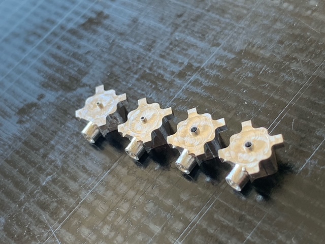
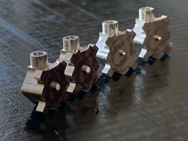
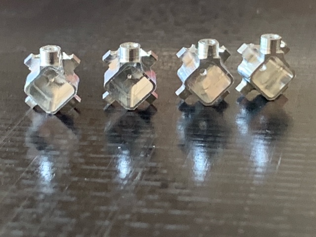
Baseplates with GRIN lens sleeves

1,201 views
Skip to first unread message

demo...@gmail.com

unread,
May 8, 2021, 3:00:49 PM5/8/21
to Miniscope

Hi Everyone –

Shylo Stiteler now has baseplates with integrated GRIN lens sleeves (see images attached).   Here is some info from him: 

He has versions to fit 1mm or 0.5 mm GRIN lenses.  Current versions are 5mm total height with a 4mm pocket.  Standoff wall thickness is 0.25 mm.  The next round will be done on a per order basis. After that the parts will have a .5mm bottom wall thickness with a .5mm standoff. 

You can contact him here to order or ask questions:

 https://miniscopeparts.com/

We have been testing early versions of these out and they work great.  Our general workflow is:

1. Attach GRIN lens holder to calibration slide or slide with fluorescent-labeled neurons.  You can see Ariel Burman’s lens holder here: https://github.com/moormanlab/miniscope-goodies

2. Put GRIN lens in lens holder.

3. Attach baseplate to Miniscope and mount scope+baseplate on stereotaxic arm.  The scope holder we use is at our github.

4. Lower scope+baseplate onto GRIN lens until image from slide comes into focus.

5. Glue GRIN lens to baseplate (just using superglue for now, but probably can use something better in the future).

6. Coat lens surface with silk+GCaMP virus as in Jackman et al., 2018.

7. Implant baseplate+lens+silk/virus in one surgery.  The baseplate holder that Ariel designed for surgery is at our github.

We’re still refining, but we now can get baseplated mice with virus at the end of a GRIN lens in a single ~3 hour surgery.

I’m sure there are still adjustments to be made, but I think we’re basically at single-surgery implants from now on.  Let us know if you have suggestions, ideas, thoughts, etc.

Thanks so much to Shylo for developing these new baseplates and letting us test them out, thanks to Daniel for the input and suggestions, and thanks to Ariel for helping with development.

- David

image0.jpeg
image1.jpeg
image3.jpeg
image2.jpeg

Brice delaCrompe

unread,
May 10, 2021, 3:03:44 AM5/10/21
to demo...@gmail.com, Miniscope
Dear David,
Thanks a lot for sharing the information and the workflow. Will be really useful!

Have a nice day,
Brice

--
You received this message because you are subscribed to the Google Groups "Miniscope" group.
To unsubscribe from this group and stop receiving emails from it, send an email to miniscope+...@googlegroups.com.
To view this discussion on the web visit https://groups.google.com/d/msgid/miniscope/6ebbbba0-a3a0-407f-881c-764d3e273926n%40googlegroups.com.


--
Brice de la Crompe, PhD
Optophysiology - Optogenetics and Neurophysiology
Albert Ludwigs University
Albertstr. 23
79104 Freiburg
Germany

Daniel Aharoni

unread,
May 10, 2021, 6:11:36 PM5/10/21
to Miniscope
Thanks for sharing this David and thanks to Shylo for pushing this project forward. We haven't had a chance to test these ourselves but they look great!

Andrew Hardaway

unread,
Nov 16, 2022, 10:30:55 AM11/16/22
to Miniscope
Hi,

How are these integrated baseplates going for people? Has anyone else used them and can share their experience with them?

best,

Andrew Hardaway

David Moorman

unread,
Nov 16, 2022, 10:45:58 PM11/16/22
to Andrew Hardaway, Miniscope
Hi Andrew - 

We're actively working on this now.  We had pretty good success in getting an initial batch working - managed to get some data from CA1 as a pilot test.  But we're now dealing with little issues like getting the GRIN lens positioned over a target in our lens holder to ensure good imaging resolution when fixing the lens to the baseplate.  Also making sure we can get the lens into the baseplate without scratching it, etc.  Little things like that.  I think we're close to getting it pretty turnkey, but there's still a little testing to do.

Happy to chat further and explain what we're doing in detail - either in the group or via email.  It would also be great to hear what other people are doing to see how we can extract best practices.

- David

Eric Melonakos

unread,
Nov 17, 2022, 11:06:04 PM11/17/22
to Miniscope
Hi all,

I've also been using these baseplates, with 0.6 mm lenses. I'm sold on them! I struggled to get good images for a long time, and the ability to image as I'm implanting with these baseplates has been huge for me (not to mention it reduces the number of procedures from 3 to 2).

I've attached a protocol I wrote for epoxying the lens into the baseplate. Some thoughts:
  1. The lens needs to be epoxied into the baseplate before the surgery. Therefore, you really are just trading the ability to image as you cement the baseplate over the lens for the ability to image as you cement the lens in place in the brain. This means that you should be careful that, when you epoxy the baseplate to the GRIN lens, your test slide is in focus in the middle of the EWL range. I use some of the 3D printed parts David was kind enough to share above to hold the lens (modified for a 0.6 mm lens) and miniscope/baseplate.
  2. Since you can image as you lower the lens into the brain, there is a temptation to chase fluorescent neurons. This can be a double edged sword: I ended up 1 mm or so deeper in one of my rats than I intended. During the surgery, I saw some static fluorescence. However, when I imaged the animal awake a week or so later, it was dark. My hypothesis, supported by the histology, is that I compressed the tissue, and as it relaxed around the lens, the fluorescent neurons rose above the bottom of the lens.
  3. As David mentioned, it can be tricky to get the lens into the baseplate. The fit is pretty snug, so as you're using a stereotax to lower the baseplate onto the lens, go slowly. I put my test slide on a plastic pipette box. That way if I catch the edge of the lens as I'm lowering the baseplate, the lens pushes on the top of the box and I can see the plastic bend a little. If it was rigid, it would probably chip the lens. It usually takes a number of tries before it goes down over the lens, but it's not too bad.
Hope this helps,

Eric
Epoxying_the_GRIN_lens_to_the_baseplate_09212022.docx

Andrew Hardaway

unread,
Nov 18, 2022, 10:08:50 AM11/18/22
to Eric Melonakos, Miniscope
David and Eric,

Wow this is terrific feedback and I'm so excited to try it. Thank you guys for sharing your protocol with me.

Couple of questions:

1) Any GCaMP or GFP fluorescent sample will do to place under the lens as a test?

2) Where have you guys been imaging with this approach?

Based on how many of the integrated lenses Inscopix is selling it seems like there might be a market in this community for sale/purchase of the integrated lenses for the V4.

best,

Andrew


--
J, Andrew Hardaway
Assistant Professor
Department of Psychiatry & Behavioral Neurobiology
University of Alabama at Birmingham School of Medicine
he/him/his

Eric Melonakos

unread,
Nov 19, 2022, 4:01:38 PM11/19/22
to Miniscope
  1. I use this test target from Thor Labs (https://www.thorlabs.com/thorproduct.cfm?partnumber=R1DS1P) with a little piece of fluorescent green flagging tape (https://ca.vwr.com/store/product/en/8545290/flagging-tape-nmc-national-marker-company) taped to the back of the test target. You might be able to use a brain slice or something on a slide, too, but you'd want to make sure the distance from the GRIN lens to the fluorescent neurons was similar to the distance you'll have in live animals.
  2. I've tried motor cortex and the prelimbic cortex, both in rats. I started with motor cortex and had a very low success rate, and then switched to prelimbic cortex and have had more success. It may just be that I've gotten better at it, though, as I was new to calcium imaging and miniscopes.

David Moorman

unread,
Nov 19, 2022, 9:54:48 PM11/19/22
to Miniscope
Thanks for sharing everything Eric - this is super helpful.  I shared your protocol with our lab and I think we'll try the plastic box idea when attaching the lens - great idea.

We got similar test targets from Thorlabs, though we got negative not positive tests.  In retrospect we probably should have gotten positives, though I think the negatives are working OK.  

Our issue has been getting the lens holder precisely positioned over the target so that the GRIN lens is aligned.  In the interim while we figure that out, we've been using GFP-labeled neuron slides that have really bright and dense fluorescence so that anywhere you put your lens you're going to see neurons.  That works well.  You can also use fluorescent beads, which should also be evenly-distributed throughout a slide.

So far we've only imaged in CA1, just to test things out, and it worked.  But we'll be trying to get this working in hypothalamus and PFC soon.  We're going to try prism lenses in PFC.  Getting those into the baseplates before implanting involves a slightly different setup, but Ariel Burman (who was the one in my lab who set all of this up) got it working with some additional parts.  Happy to discuss that with anyone interested.

Anyone - please send suggestions for improvements or changes.  It would be great if we could get this to be 100% reliable and easy for everyone. 


Andrew Hardaway

unread,
Nov 20, 2022, 10:15:21 AM11/20/22
to David Moorman, Miniscope
Thank you David for the helpful feedback! We are very excited to try it out. 

Hao Li

unread,
Apr 3, 2025, 4:42:38 PMApr 3
to Miniscope
Hi everyone, 
We are new to Miniscope and came across this intergrated baseplate, which looks amazing. I was wondering how they are working out for you and particularly how to position the lens to the working distance. 
Thanks in advance,
Hao

David Moorman

unread,
Apr 3, 2025, 5:48:27 PMApr 3
to Hao Li, Miniscope
Hi Hao and all - 

Short answer is that they're still working well for us.  We've been able to do some good imaging in a couple of different brain areas with them. 

Positioning them is pretty easy.  We set the scope at the 0 position and mount the baseplate on the scope.  Then we image as we're lowering the baseplate onto the lens.  When we get good resolution on our image, we stop and glue it.  It's relatively qualitative, but it seems to work OK.  There is some debate in the lab about whether we should set the scope at the highest position (thus maximizing working distance away from the lens face), and we haven't decided what's best.  Happy to take suggestions/opinions on this.

We have lens and slide holders on our GitHub (https://github.com/moormanlab/miniscope-goodies/tree/main/3Dmodels/stls), though we haven't updated them in a while, and I'm sure they can be tweaked to better suit your needs.

A couple of caveats based on our experience so far:

1.  They are easier to use with 1 mm lenses than 0.5 mm lenses.  The 0.5mm lenses are so fragile that trying to get them into a sleeve that fits them often breaks them.  So for smaller diameter lenses we have been using the traditional baseplates.  Getting 1 mm lenses in the sleeve is a bit easier.

2.  We aren't using the silk+virus strategy - we're just injecting the virus as normal.  Sometimes we do 2 surgeries - 1st for the virus and 2nd for the lens.  We've been told that putting the lens in with the virus in the same surgery can displace the virus, which is an issue when you're targeting a small brain region.  Not sure how often this happens, but if we're able to take our time, we've been doing a bunch of virus surgeries and then lens implants after just in case.

I'll let other folks chime in.  Happy to answer other questions.  We've been spending a long time piloting this out but I think we're getting pretty consistent results at this point (thanks in part to everyone's feedback on this email group).

- David

Hao Li

unread,
Apr 11, 2025, 12:08:45 PMApr 11
to David Moorman, Miniscope
Hi David,

Thanks so much for the detailed response! Really appreciate you sharing your experience. It’s great to hear the sleeves have been working well for you and that you’re getting good imaging consistently. We are mostly working with 0.5/0.6mm lenses and are excited to try it!

Thanks,
Hao
--
Thanks,

Hao

Ed Hayter

unread,
Apr 11, 2025, 12:08:58 PMApr 11
to Miniscope
Just to add to David's comment - For the smaller 0.5/0.6mm lenses I've had much more success holding the lens with a cannula holder and lowering it into the upside-down baseplate, the caveat being you can't image as you go. I've found that as long as the lens protudes slightly into the baseplate (which is also helpful for cleaning between uses) you end up with a decent working distance which covers imaging right next to the lens face, to a couple hundred microns away. Again, not perfect but works well here. To get the distance right you can print a simple chuck to hold the baseplate and stop the lens just as it sticks out of the hole. Happy to share pictures if anything is unclear!

Best,
Ed
Reply all
Reply to author
Forward
0 new messages