Unable to see active cells
391 views
Skip to first unread message

Longwen Huang
Jul 1, 2022, 3:33:23 AM7/1/22
to Miniscope
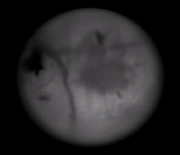
Hi, we're using miniscope v4 to record from a deep structure (~2mm) in mice, but we got some issues. We saw cells initially, but they didn't change activity during behaviors (high-titer1.avi). They disaapeared 2 weeks later (high-titer2.avi). So we next lowered the titer and confirmed good shapes of cells in slices (low-titer-slice.tif). But during in vivo imaging, we only saw some glow at high (>50) LED intensity (low-titer1.avi). We electro tuned the focal plane of the lens, but cells never showed up. Any advice? Thanks!
Longwen Huang
Jul 1, 2022, 3:33:50 AM7/1/22
to Miniscope
Some information that might
be useful: we used a go-foton lens with ~4-6mm length. Before we implanted the
lens, we used a pipette connected to a vacumm to suck up tissues 1.6mm from the
brain surface. Then the grin lens was implanted 1.9mm from the brain surface.
When we mounted the baseplate, we first focused on the lens surface, and then
moved 100-200um above. All surgeries were done 3 weeks after viral injection.

Hannah Wirtshafter
Jul 1, 2022, 2:32:26 PM7/1/22
to Miniscope
Did you actually put your videos through post processing? I thought I had the same issue when I started but after post processing they were clearly firing
Longwen Huang
Jul 3, 2022, 9:16:29 PM7/3/22
to Miniscope
Thanks for your reply. We have used minian to process the video, but we still couldn't see any firing cells in the post-processed video.

Federico Sangiuliano
Jul 4, 2022, 1:41:41 AM7/4/22
to Miniscope
Can you upload the video of "high-titer1.png"? It looks like you have some neurons in your field of view but I can't really tell from a picture

xiaoyu peng
Jul 4, 2022, 8:02:09 AM7/4/22
to Longwen Huang, Miniscope
Hi, Longwen,
If you image soon after lens implant surgery, the neurons may be there, but their function may not fully recover yet, they may be like in stroke. In my hand, In cortex imaging, they seem to "freeze". They may need weeks to get active. I am not sure if this applies to your brain region. I end up doing virus injection and lens implantation or cranio window in the same surgery, so I waited in total shorter time. Your high-titer-2 field of view seems quite different from high-titer-1, could you also check baseplate position? Otherwise, the tissue may just regenerate a bunch. It seems that you still have some signal in high-titer2, but not in focus, not sure if you can focus down more. What virus serotype are you using? I like AAV1.
Best,
Xiaoyu
On Sun, Jul 3, 2022 at 7:16 PM Longwen Huang <huangl...@gmail.com> wrote:
Thanks for your reply. We have used minian to process the video, but we still couldn't see any firing cells in the post-processed video.Did you actually put your videos through post processing? I thought I had the same issue when I started but after post processing they were clearly firing
--
You received this message because you are subscribed to the Google Groups "Miniscope" group.
To unsubscribe from this group and stop receiving emails from it, send an email to miniscope+...@googlegroups.com.
To view this discussion on the web visit https://groups.google.com/d/msgid/miniscope/c5b482ad-866e-49e5-8bf0-bc8534916813n%40googlegroups.com.
LH
Jul 5, 2022, 4:18:38 AM7/5/22
to Miniscope
Hi Xiaoyu, thanks for your information. We were pretty sure that the baseplate was held very tightly. Also it looked like that the same blood vessle were present in the left of both high-titer-1 and high-titer-2, but the cells were gone and we couldn't see them within the electro-tunable focal range. We also saw lots of auto-fluorescence - but again no cells - around the grin lens track in the slices after sacrificing the animal.
Also, we're using AAV2/9.

Nate Klett
Jan 13, 2023, 6:13:31 AM1/13/23
to Miniscope
This is very interesting. Could you elaborate on this? Is it expected to see cells flashing during base-plating with the raw videos? Or will this only become obvious after processing the videos? Cheers.

David Protter
Jan 23, 2023, 12:14:35 PM1/23/23
to Miniscope
Depends on the brain region if you see it during baseplating under anesthesia. For AAV based expression, I wait 3-4 weeks before baseplating and wake the animal up for a 5-10 minute "cell-check" imaging session just in an open field box. If I dont clearly see active units then in the df/f mode, I basically never have seen active units down the line. Sometimes I will see activity in the raw video (not in df/f) if I have a few particularly active cells, but its much harder to notice by eye most of the time.
(ive been using jGCaMP7f, which is pretty comparable to 6f in terms of real world SNR, i hear.)
I will say it is quite hard for me to estimate how many units i'm looking at with the daq streaming visualization vs when I used an inscopix scope (just different scaling on the streamed video), but its usually very obvious if an animal does or doesnt have activity.
I will say it is quite hard for me to estimate how many units i'm looking at with the daq streaming visualization vs when I used an inscopix scope (just different scaling on the streamed video), but its usually very obvious if an animal does or doesnt have activity.
Reply all
Reply to author
Forward
0 new messages