EmbryoPhysics81:: Embryo Physics Course: Diatom morphogenesis
Richard Gordon
Gordon, R. & R.W. Drum (1994). The chemical basis for diatom morphogenesis. Int. Rev. Cytol. 150, 243-372, 421-422. (80MB file, available on request)
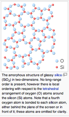
in terms of the radial distribution function, as determined by x-ray diffraction and other methods, and concluded that there were simply no observations on fresh or live diatom shells, which would be the most relevant. I’d guess that such work still has to be done.
Borowitzka, M.A. & B.E. Volcani (1978). The polymorphic diatom Phaeodactylum tricornatum: ultrastructure of its morphotypes. J. Phycol. 14(1), 10-21.
are on p.249 of Gordon & Drum (1994). The role of organic matter is in dispute. See:
Gordon, R., D. Losic, M.A. Tiffany, S.S. Nagy & F.A.S. Sterrenburg (2009). The Glass Menagerie: diatoms for novel applications in nanotechnology [invited]. Trends in Biotechnology 27(2), 116-127. (attached)
What the computer simulations suggest is that, given a nucleating center (linear for pennate diatioms, and a small ring for centric diatoms), it is plausible that there is no relevant structure within the silicalemma. Empirical evidence would be nice to have. Conceptually, I would argue that such structure would be deemed a prepattern, whose pattern would then need to be explained. If precipitation of silica proves an adequate explanation for the gross features of the pattern, then why not apply Occam’s razor? Of course, observation trumps a priori reasoning, which is why I call for more observation.
Chaos theory and complexity theory may have their place here, but would not explain the constant features that are shared by daughter cells and bigger clones. Thus they are not easily brought in as an explanation of why and how there are 100,000 morphologically distinct species.
I’m getting mixed messages regarding further interest in diatom morphogenesis and morphogenesis of other single cell organisms, such as ciliates: low attendance today and 3-4 people who couldn’t make it asking me to repeat it. I’ve decided to repeat the talk later and get back to vertebrates next week:
Dick Gordon Hierarchical Genome: The Whole Megillah in 1 Hour May 12, 2010
Dick Gordon Condensed repeat of diatom morphogenesis, + motility (new), in 1 hour June 9, 2010
The latter date is adjustable on request.
Thanks.
Yours, -Dick
From: Stephen M. Levin <sml...@biotensegrity.com>
Date: Wed, May 5, 2010 at 5:56 PM
Subject: Re: Embryo Physics Course: Diatom morphogenesis
To: Richard Gordon <dickgo...@gmail.com>


Tuesday, May 4, 2010 8:15 AM from Winnipeg, MB, Canada
Dear Friends,
Tomorrow (Wednesday May 5) I’ll be talking on single cell
morphogenesis whose ornateness would seem to exceed that of many
multicellular organisms, in:
Embryo Physics Course, http://embryophysics.org, most Wednesdays at
2pm Pacific Time, held online at
http://slurl.com/secondlife/Silver%20Bog/84/32/60.
If you need help in getting started in Second Life®
(http://secondlife.com), give me or William R. Buckley a holler:
gor...@cc.umanitoba.ca
W...@wrbuckley.com
Thanks.
Yours, -Dick Gordon
--
Dr. Richard Gordon, Professor, Radiology, University of Manitoba
GA216, HSC, 820 Sherbrook Street, Winnipeg R3A 1R9 Canada
E-mail: gor...@cc.umanitoba.ca, Skype: DickGordonCan, Second Life:
Paleo Darwin, Cell: 1-(204) 995-7125
Embryo Physics Course: http://embryophysics.org/;
http://bookswithwings.ca; Adjunct Scientist: TRLabs, http://www.win.trlabs.ca/
http://www.umanitoba.ca/faculties/medicine/radiology/stafflist/rgordon.html
Affiliate, Institute of Industrial Mathematical Sciences (IIMS),
http://www.umanitoba.ca/institutes/iims/
Principal Scientific Advisor, EvoGrid: http://www.evogrid.org
--
Dr. Richard Gordon, Professor, Radiology, University of Manitoba GA216, HSC, 820 Sherbrook Street, Winnipeg R3A 1R9 Canada
E-mail: gor...@cc.umanitoba.ca, Skype: DickGordonCan, Second Life: Paleo Darwin, Cell: 1-(204) 995-7125
Embryo Physics Course: http://embryophysics.org/;
http://bookswithwings.ca; Adjunct Scientist: TRLabs, http://www.win.trlabs.ca/
http://www.umanitoba.ca/faculties/medicine/radiology/stafflist/rgordon.html
Affiliate, Institute of Industrial Mathematical Sciences (IIMS), http://www.umanitoba.ca/institutes/iims/
Principal Scientific Advisor, EvoGrid: http://www.evogrid.org
You received this message because you are subscribed to the Google Groups "EmbryoPhysics" group.
To post to this group, send email to embryo...@googlegroups.com.
To unsubscribe from this group, send email to embryophysic...@googlegroups.com.
For more options, visit this group at http://groups.google.com/group/embryophysics?hl=en.
