multiple phase fitting and electron diffraction
125 views
Skip to first unread message

laura
Mar 22, 2021, 7:37:42 AM3/22/21
to diffpy-users
Dear all,
I am new with PDF refinements and also I have to say that I am trying to do it with electron diffraction.
The point is that I have a sample with a SAED that seems quite amorphous with only 3 diffuse rings, so knowing what has been formed with diffraction is complicated. Now I try with PDF and that is why I am here!. However, I have some possible candidates but it is like trial-error. I have some questions: when making a fitting with multiple phases (3 or more), what is the procedure to follow?: I do enter the phases together with the data but how do I assign the @ for each phase so that they are relative?.
How do you know that those candidates are correct, I mean, before you start to make a deeper fit?. It is simple when there is one phase because the peaks have to be in the possition but what happen when there are several phases and still do not know the quantities for each phase?.
And finally, what parameters are important when working with electron diffraction in PDFgui?,
Thank you so much in advance for your help!
Laura Pascual

Simon Billinge
Mar 24, 2021, 8:22:11 AM3/24/21
to diffpy...@googlegroups.com
Hi Laura,
These are complicated questions that it is hard to answer on a thread like this. It really depends on the details of the scientific question you are after.
If your signal is amorphous it won't contain much information. From an information theoretic point of view, if you have 3 separated peaks in your PDF you have roughly 9 bytes of information and it only makes sense to fit models that are constrained to have < 9 numbers that specify them. So try and come up with simple questions/hypotheses that you can ask to your data that can be tested and build models to test them.
If you are fitting multiple phases to an amorphous signal, what do you mean? You think that there are 3 different glasses in the region where your beam hit? Without more info I can't say, but it seems unlikely. If it was true you would need 3 numbers to specify the quantities of each phase, so you already used up 3 of your 9 numbers, so don't let too many parameters vary in the models themselves.
OK that is just some general thoughts to maybe help you to get started. These are not issues specific to PDF but good to think about.
S
--
You received this message because you are subscribed to the Google Groups "diffpy-users" group.
To unsubscribe from this group and stop receiving emails from it, send an email to diffpy-users...@googlegroups.com.
To view this discussion on the web visit https://groups.google.com/d/msgid/diffpy-users/052c75b6-a00f-4845-a927-894e3ef8bc60n%40googlegroups.com.
--
Simon Billinge
Professor, Columbia University
Physicist, Brookhaven National Laboratory
Laura
Mar 24, 2021, 6:25:26 PM3/24/21
to diffpy...@googlegroups.com
Dear Simon,
I really appreciate your answer. Sorry for not posting the question correctly, or with little data. The point is that I start from a well crystallized phase (perovskite) that, when put into electrolyte (acidic media), loses cations and becomes amorphous, leaving Ir, O and only a small fraction of the other atoms.
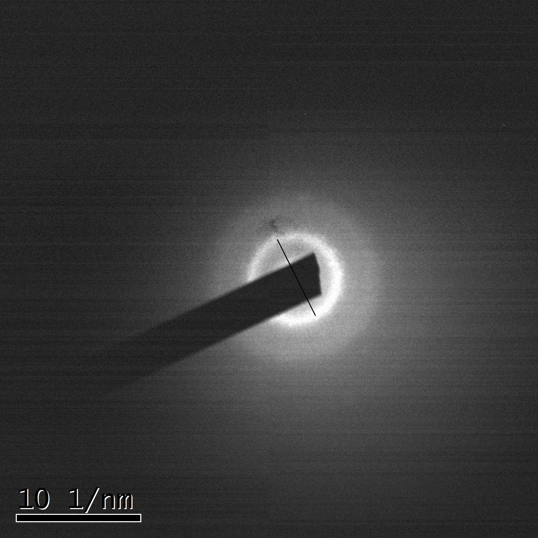
In the SAED there are three diffuse rings so the domains must be small. The resulting phase is quite active in the reaction, so, It would be interesting to know what happens here. I can't identify
the structure with just those three rings either, so I go blind. The subject with several phases comes from this paper: E. Willinger et al. J. Am. Chem. Soc. 2017, 139, 34, 12093–12101 , where they identify in the IrOx
the presence of hollandite together with rutile-type IrO2 through electron diffraction and PDF analysis. In my case I am supposing that the starting structure remains with disorder and some related candidates may appear, for example IrO2 and/or Ir metallic,.but I do not know if this is possible or the problem is so complex and not able to fit it. I send you the SAED pattern and also the PDF file obtained from it.
I hope this may clarify what I mean.
Thanks a lot for your time and your consideration,
Laura
To view this discussion on the web visit https://groups.google.com/d/msgid/diffpy-users/CANugsUEB2LVuzLcK2VJJ-h7zozUm5qK7XWrLzYORJp3bL3hquw%40mail.gmail.com.

GiHan Kwon
Dec 8, 2021, 10:25:45 PM12/8/21
to diffpy-users
Hi Laura
I am also studying amorphous IrOx thin film on electrode surface for ex-situ and in-situ electrochemical PDF measurement. I knew Willinger paper you mentioned. In her paper peak position is not identical although the same sample was used. I guess it was from a kind of electron beam damage which is much serious than X-ray beam damage.
my data fitting in the paper (https://pubs.acs.org/doi/10.1021/acscatal.1c00818) was not really great due to an amorphous character. thus I used two fragments (mother model and fragment from mother model). usually, a raw structure was obtained from ICSD database and the mother model was made with discovery studio or any 3D software like Mercury or Vesta too. Actually, a raw structure is not fit well to experimental data. thus I used diffpy-CMI to move each atom a little bit with different ADP on each atom and diffpy-CMI did also fit G(r). after obtaining mother model structure by diffpy-CMI, with my own code I calculate I(q), S(q), F(q), and G(r) to check patterns with especially I(q). Fitting experimental I(q) is the best way. Usually, protein crystallographers do. Once these procedures are done, I gave different weights on each model (mother model and fragment) to fit experimental data. Regarding beam damage by electron or X-ray, some of iridium samples are so sensitive. you will find alteration of the structure by electron and X-ray beam in the SI too.
please let me know if you have any questions.
Thank you,
Gihan
2021년 3월 24일 수요일 오후 6시 25분 26초 UTC-4에 laura님이 작성:
Reply all
Reply to author
Forward
0 new messages