segmentation using phase and fluorescence images missin signal
198 views
Skip to first unread message

toco...@jhmi.edu
Feb 26, 2021, 7:21:21 AM2/26/21
to MicrobeJ Project
Hi,
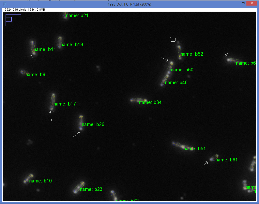
I am trying to measure fluorescence intensity along the medial axis of rod shaped bacteria to monitor protein localization. A significant portion of the protein localizes to the poles in wild type bacteria. I have tried using a phase image to do segmentation however, in the majority of cases, the resulting area defined partly, or in rare cases entirely excludes the polar puncta (see attached image, arrows). I have tried to extend the region of interest using the extension option under profile but that did not help. I have also tried using the fluorescence image to do segmentation however, since the bacteria often have two polar puncta (wild type bacteria) or multiple distinct puncta (mutant bacteria), the segmentation treats these as individual cells. I have tried to link them by altering the z score which helps in a couple cases but not for the majority of bacteria.
We are currently trying to stain the bacteria more uniformly with a second dye but we would like to be able to analyze this large data set that we already have.
Thanks,
Tamara
Tamara

ellenmq
May 5, 2021, 2:42:59 AM5/5/21
to MicrobeJ Project
Hi Tamara,
Do you also have phase images to go with these fluorescence images that could be used for segmentation? If so, I think that would resolve this issue. You would process the phase and fluorescence images together, but use the phase image to detect the whole bacteria outline, then the maxima feature to detect fluorescent foci.
Do you also have phase images to go with these fluorescence images that could be used for segmentation? If so, I think that would resolve this issue. You would process the phase and fluorescence images together, but use the phase image to detect the whole bacteria outline, then the maxima feature to detect fluorescent foci.

Jordan Barrows
Jan 14, 2022, 11:32:49 AM1/14/22
to MicrobeJ Project
Hi Tamara,
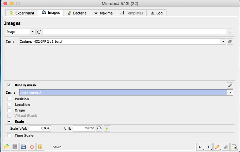
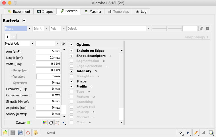
I've been thinking more about this, and I'm wondering if you might be able to generate binary images in FIJI first (https://imagej.nih.gov/ij/docs/guide/146-29.html), and then upload that image into MicrobeJ as a binary and use it to perform the segmentation (see attached screenshots, and feel free to disregard the other settings), proceeding then to use your fluorescence image to detect maxima/fluorescence intensity along the cell(s).
Let me know if this helps.
Jordan
Reply all
Reply to author
Forward
0 new messages