A probabilistic atlas of the human thalamic nuclei

Asa Borzabadi

Glasser, Matt
That might be a better question for the FreeSurfer list. We have not compiled these new atlases for the HCP data at this point.
Matt.
--
You received this message because you are subscribed to the Google Groups "HCP-Users" group.
To unsubscribe from this group and stop receiving emails from it, send an email to
hcp-users+...@humanconnectome.org.
To view this discussion on the web visit
https://groups.google.com/a/humanconnectome.org/d/msgid/hcp-users/CA%2Bs6gv%3Dq6RoY2zF2UOEBP77LQEHOq%2BmpQTSjxH%2BDXV9GVskL8Q%40mail.gmail.com.
The materials in this message are private and may contain Protected Healthcare Information or other information of a sensitive nature. If you are not the intended recipient, be advised that any unauthorized use, disclosure, copying or the taking of any action in reliance on the contents of this information is strictly prohibited. If you have received this email in error, please immediately notify the sender via telephone or return mail.
Asa Borzabadi
To view this discussion on the web visit https://groups.google.com/a/humanconnectome.org/d/msgid/hcp-users/B0CC39AC-6600-4ABC-962D-7C5AB8B05CD4%40wustl.edu.

Harms, Michael
Is this a volume (mgz) file that FS spits out for each subject, which contains a set of thalamic nuclei labels customized to the anatomy of each subject?
If so, adding the relevant image volume name to this ‘for’ loop in FreeSurfer2CaretConvertAndRegisterNonlinear.sh may be all that you need to do:
#Convert FreeSurfer Volumes
for Image in wmparc aparc.a2009s+aseg aparc+aseg ; do
if [ -e "$FreeSurferFolder"/mri/"$Image".mgz ] ; then
mri_convert -rt nearest -rl "$T1wFolder"/"$T1wImage".nii.gz "$FreeSurferFolder"/mri/"$Image".mgz "$T1wFolder"/"$Image"_1mm.nii.gz
applywarp --rel --interp=nn -i "$T1wFolder"/"$Image"_1mm.nii.gz -r "$AtlasSpaceFolder"/"$AtlasSpaceT1wImage" --premat=$FSLDIR/etc/flirtsch/ident.mat -o "$T1wFolder"/"$Image".nii.gz
applywarp --rel --interp=nn -i "$T1wFolder"/"$Image"_1mm.nii.gz -r "$AtlasSpaceFolder"/"$AtlasSpaceT1wImage" -w "$AtlasTransform" -o "$AtlasSpaceFolder"/"$Image".nii.gz
${CARET7DIR}/wb_command -volume-label-import "$T1wFolder"/"$Image".nii.gz "$FreeSurferLabels" "$T1wFolder"/"$Image".nii.gz -drop-unused-labels
${CARET7DIR}/wb_command -volume-label-import "$AtlasSpaceFolder"/"$Image".nii.gz "$FreeSurferLabels" "$AtlasSpaceFolder"/"$Image".nii.gz -drop-unused-labels
fi
done
cheers,
-MH
--
Michael Harms, Ph.D.
-----------------------------------------------------------
Associate Professor of Psychiatry
Washington University School of Medicine
Department of Psychiatry, Box 8134
660 South Euclid Ave. Tel: 314-747-6173
St. Louis, MO 63110 Email: mha...@wustl.edu
From: Asa Borzabadi <asa.bo...@gmail.com>
Reply-To: "hcp-...@humanconnectome.org" <hcp-...@humanconnectome.org>
Date: Monday, May 2, 2022 at 2:36 PM
To: "hcp-...@humanconnectome.org" <hcp-...@humanconnectome.org>
Subject: Re: [hcp-users] A probabilistic atlas of the human thalamic nuclei
|
* External Email - Caution * |
To view this discussion on the web visit https://groups.google.com/a/humanconnectome.org/d/msgid/hcp-users/CA%2Bs6gvnAd%2B98uaBMVGDkxKX7GXWsPQGWqfjdQp1cRgertUyq3A%40mail.gmail.com.
Asa Borzabadi
mri_convert -rt nearest -rl ${SubjectDir}/${Subjectid}/T1w/T1w_acpc_dc_restore.nii.gz ${SubjectDir}/${Subjectid}/T1w/${Subjectid}/mri/ThalamicNuclei.v13.T1.FSvoxelSpace.mgz ThalamicNuclei.v13.T1.FSvoxelSpace.nii.gzapplywarp --rel --interp=nn -i ThalamicNuclei.v13.T1.FSvoxelSpace.nii.gz -r ${SubjectDir}/${Subjectid}/MNINonLinear/T1w_restore.nii.gz -w ${SubjectDir}/${Subjectid}/MNINonLinear/xfms/acpc_dc2standard -o ThalamicNuclei.v13.T1.FSvoxelSpace_native.nii.gzapplywarp --interp=nn -i ThalamicNuclei.v13.T1.FSvoxelSpace_native.nii.gz -r ${SubjectDir}/${Subjectid}/ MNINonLinear/ROIs/Atlas_ROIs.2.nii.gz -o ThalamicNuclei.v13.T1.FSvoxelSpace_Atlas_2mm.nii.gzwb_command -volume-label-import ThalamicNuclei.v13.T1.FSvoxelSpace_Atlas_2mm.nii.gz FreeSurferColorLUT.txt ThalamicNuclei.v13.T1.FSvoxelSpace_Atlas_2mm.nii.gz -drop-unused-labels
To view this discussion on the web visit https://groups.google.com/a/humanconnectome.org/d/msgid/hcp-users/92677B55-A615-4CD4-8C52-10AA50E0ECA8%40wustl.edu.

Coalson, Timothy Scott (S&T-Student)
Sent: Monday, August 1, 2022 9:06 AM
To: hcp-...@humanconnectome.org <hcp-...@humanconnectome.org>
Subject: Re: [hcp-users] A probabilistic atlas of the human thalamic nuclei
Asa Borzabadi
To view this discussion on the web visit https://groups.google.com/a/humanconnectome.org/d/msgid/hcp-users/SN6PR01MB499121B136C9E819E268AFBC819A9%40SN6PR01MB4991.prod.exchangelabs.com.
Glasser, Matt
Is this an individual subject or an average?
Matt.
From: Asa Borzabadi <asa.bo...@gmail.com>
Reply-To: "hcp-...@humanconnectome.org" <hcp-...@humanconnectome.org>
Date: Wednesday, August 10, 2022 at 2:41 PM
To: "hcp-...@humanconnectome.org" <hcp-...@humanconnectome.org>
Subject: Re: [hcp-users] A probabilistic atlas of the human thalamic nuclei
Thank you so much for the help, and thanks for the correction.
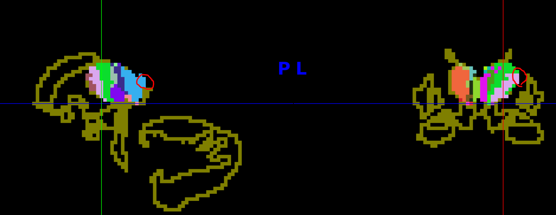
I still have a general question. When viewing the converted intra-thalamic label files (now in MNI152_2mm space) on top of the Atlas.ROIs.2.nii.gz template, there exist voxels outside the boundary of Atlas.ROIs.2.nii.gz template (their existence could possibly be explained by the fact that FreeSurfer7, which does the intra-thalamic segmentation, uses a different approach for thalamic parcellation than FreeSurfer6); in other words, there exist some voxels that are not defined in the standard subthalamic space of HCP.
(the figure is attached for better illustration, the layout of the template and the resulted intra-thalamic segmentation are plotted, some inconsistent voxels are pointed to with red circles)

To view this discussion on the web visit https://groups.google.com/a/humanconnectome.org/d/msgid/hcp-users/CA%2Bs6gv%3DyxU15NTFjxT9e-t89riuZAR-BTfgM7FiUkZv4dgM%3DLQ%40mail.gmail.com.
Asa Borzabadi
To view this discussion on the web visit https://groups.google.com/a/humanconnectome.org/d/msgid/hcp-users/0C8111AD-95B4-4C88-A8FD-8BCCA7D9CE19%40wustl.edu.
Glasser, Matt
Is it possible that things aren’t quite lined up in the volume? What did you use for volume registration?
Matt.
To view this discussion on the web visit https://groups.google.com/a/humanconnectome.org/d/msgid/hcp-users/CA%2Bs6gvmyMLUNxrMTrLTv9qmNigJcYBjkSthKgUj%2BHU7X94BGDw%40mail.gmail.com.
Harms, Michael
Isn’t this fairly common? IIRC, nothing prevents the FS subcortical labels in an individual from mapping outside of the CIFTI atlas labels during the computation of the registration to MNINonLinear space.
--
Michael Harms, Ph.D.
-----------------------------------------------------------
Associate Professor of Psychiatry
Washington University School of Medicine
Department of Psychiatry, Box 8134
660 South Euclid Ave. Tel: 314-747-6173
St. Louis, MO 63110 Email: mha...@wustl.edu
To view this discussion on the web visit https://groups.google.com/a/humanconnectome.org/d/msgid/hcp-users/CC109575-2B2D-4354-8F6D-ABEF661F255A%40wustl.edu.
Glasser, Matt
It shouldn’t be off by much.
Matt.
To view this discussion on the web visit https://groups.google.com/a/humanconnectome.org/d/msgid/hcp-users/27C1994E-CD9A-4D13-AB8F-607026D9F1BA%40wustl.edu.
Harms, Michael
But it happens to at least some extent with some regularly (because the MNINonLinear registration is not constrained/informed by the FS subcortical labels). In the provided example image, the “spill-over” outside of the CIFTI atlas indeed wasn’t much.
To view this discussion on the web visit https://groups.google.com/a/humanconnectome.org/d/msgid/hcp-users/0A09413E-4B92-4B73-A727-1F3E06C6F0D6%40wustl.edu.
Asa Borzabadi
To view this discussion on the web visit https://groups.google.com/a/humanconnectome.org/d/msgid/hcp-users/8BA8E1DA-0901-471C-BAEE-C274FA791CDC%40wustl.edu.
Glasser, Matt
You can certainly analyze data within individual subject defined parcels in individuals, but I take it you want to do a group analysis, or you would have just done that. I think the question that we are trying to address is whether this new thalamic parcellation on average is including voxels that were not previously included as thalamic greymatter voxels on average by FreeSurfer (and hence are not in the CIFTI standard space) or if this is simply expected small imperfections of non-linear volume-based alignment from subject to subject. The best way to address that would be to take a large group of non-linearly aligned subjects with that new thalamic parcellation and take the mode of their parcellations and see if there is a difference with the current CIFTI grayordinates space. If not, it would be sufficient to use parcel constrained resampling between the group and individuals even with minor misalignments (as the resampling will pull data from neighboring slightly misaligned voxels into the group average locations). If there is a difference, that would suggest a need to modify the CIFTI grayordinates space.
To view this discussion on the web visit https://groups.google.com/a/humanconnectome.org/d/msgid/hcp-users/CA%2Bs6gvkWfUP_AfJmgXMz493UCTKOeDbmaCmFLWxMDGFCH4YrUg%40mail.gmail.com.
Asa Borzabadi
Glasser, Matt
Yes, but I wouldn’t expect any systemic effects and that this would be limited to a voxel from which at least some signal should be recovered when generating the CIFTI timecourse for the neighboring voxel.
To view this discussion on the web visit https://groups.google.com/a/humanconnectome.org/d/msgid/hcp-users/ee47c939-76ad-42e7-ade5-fee6ed513dd0n%40humanconnectome.org.